Microscopy day 21st May 2022
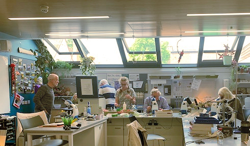
OBKA organised an Introduction to Microscopy course for beekeepers at St Clare’s School, Oxford. The course was led by member Sarah Jinks, a biology teacher at the school. We used two types of microscopes; Stereo Microscope (about x10 magnification) which gives a 3-D view and is used for dissection, and Compound Microscope which gives high magnification up to x1000 and is used for viewing prepared slides.
Ten OBKA Member attended the course and, after coffee, moved to the science laboratory to receive an introduction to the workings of each microscope. After this introduction we split into two group, one examining pollen while the other group worked on dissection and examining the anatomy of the honey bee.
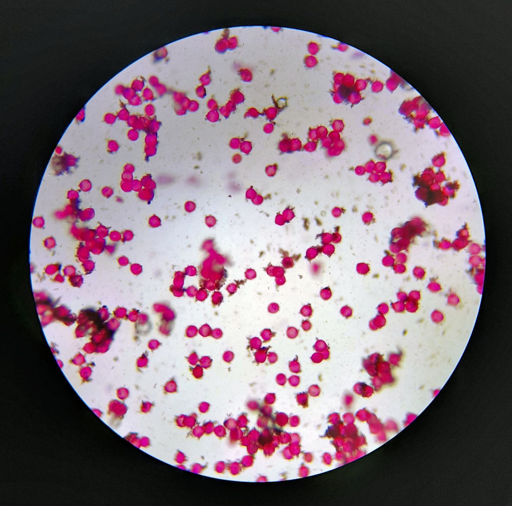
For the pollen group, the first method used was to prepare a pollen slide directly from flowers by simply dropping a staining solution on to a glass slide and gentle touching it with the flower anther to release pollen. Once a cover slip had been placed on the slide, it was then ready for viewing. Once in focus, the complex pollen shapes can be identified with the use of a pollen identification book or website. (We used “The Pollen Loads of the Honeybee” by Dorothy Hodges” which has many drawings of British pollens.) Sarah demonstrated how to recover pollen from honey using a centrifuge. The honey is first dissolved in warm water and then centrifuged for 5 minutes. This leaves the pollen at the bottom of the tube ready to put on a slide. Adding a staining agent aids viewing. We also extracted pollen from honeycomb and were able to compare a red pollen extracted from honeycomb with that collected from a horse chestnut flower. Finally, we examined varroa tray debris including wax flakes.
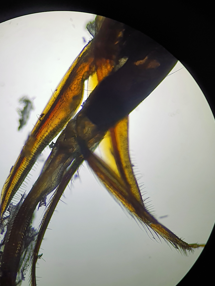
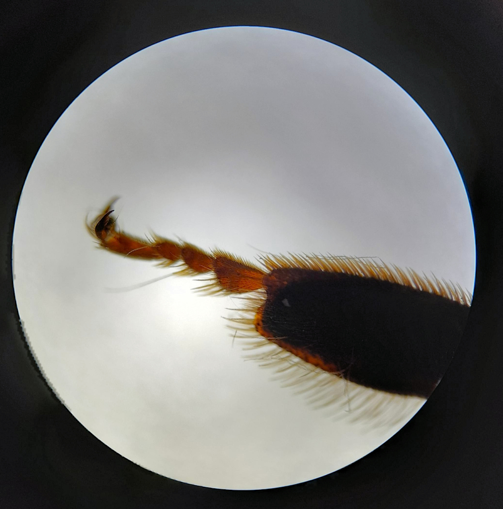
Halfway through the day we swapped groups and moved onto dissection and examination of the bee’s anatomy. The dissecting microscopy produces an enlarged image of the bee, allowing you to carefully remove external body parts such as wings, sting, venom sac, and legs and place them onto slides ready to view under the compound powerful microscope. Dissecting the external anatomy is fairly easy but removing the internal organs is very challenging.
The views of the workings of the bee’s anatomy were fantastic, especially the wing structures, legs and feet. A couple of us managed to remove the stinger with intact venom sac, and the mouth parts. One person managed to remove part of the compound eye, while others examined the trachea for evidence of acarine. We also tested for nosema, which involves grinding up the abdomen from several bees in water, applying stain and finally viewing the liquid using a high-power microscope. (NB. If used as a health check, we would use thirty bees to give a representative sample.)
Overall, everyone had a very interesting and engaging day. We would like to thank Sarah for delivering the course and to St Clare’s School for the use of their facilities.
Grateful thanks to Clare Dines and Yvonne Parks for the following images